top of page

Empowering Healthcare and life sciences with
advanced lab-on-chip technology...
Our Portfolio
Welcome to our portfolio. Here you’ll find a selection of our work. Explore our projects to learn more about what we do at Point-Of-Care Microfluidics Pvt. Ltd.


Development of FET-based Electrochemical biosensor
 AFM image of a FET-based nano biosensor |  Si-SU8 hybrid mold featuring micro-patterned structures |
|---|---|
 A model of LOC Microfluidic device on silicon platform |  A thin PDMS microfluidic device fabricated from a silicon mold using soft lithography |
 Close-up optical image of a Si-SU8 hybrid mold with micro-patterned structures |  FET-based electrochemical biosensor |
 3D simulation of ion interactions on the sensor surface |  Data collected from the FET-based electrochemical biosensor |
 Close-up AFM image of the sensor |  Electrical measurement setup for FET-based chemical sensors |
 SEM image of a sub-10nm gold nanodot array |  Optical microscope image of a gold pattern fabricated on a Si/SiO₂ substrate |
|---|---|
 AFM image of a single gold nanodot with a width of 35 nm |  AFM image of a gold nanoparticle array with a 2µm spacing |
 Confocal microscopy image of a gold nanodot array functionalized with ssDNA |  AFM image of a sub-10nm gold nanodot array with 100nm spacing |
 AFM image of a sub-10nm gold nanodot array with 100nm spacing |  Optical microscope image of a gold pattern fabricated on a Si/SiO₂ substrate |
 Photographic image of a microfluidic reactor captured using a KEYENCE microscope |  Closer view of microfluidic reactor |
|---|---|
 SEM image showcasing a nanoparticle cluster synthesized using a microfluidic reactor |  SEM image showcasing a nanoparticle cluster synthesized using a microfluidic reactor |
 Image depicting laminar flow within a Y-shaped microfluidic channel containing two immiscible liquid |  SEM image showcasing a nanoparticle cluster synthesized using a microfluidic reactor |
 SEM image showcasing a nanoparticle cluster synthesized using a microfluidic reactor |  Photographic image of a microfluidic reactor captured using a KEYENCE microscope |
 Photographic image of a microfluidic reactor captured using a KEYENCE microscope |  Image of a fabricated microfluidic reactor with tube connectors, created using soft lithography |
 Nanofluidic channels with a width of less than 100 nm |  Precisely aligned gold nanodot within an open nanochannel |
|---|---|
 Nanofluidic channels under 100 nm with perfectly aligned gold nanoelectrodes of sub-10 nm | 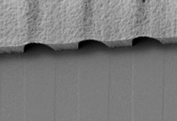 Closed Nanochannel arrays |
 Closed Nanochannel arrays |  Precisely aligned gold nanodot within a closed nanochannel |
 Precisely aligned gold nanodot within a closed nanochannel |  Three-dimensional nanofluidic structures |
Nanoelectrode array biosensors
Microfluidic reactor for colloidal and nanoparticles synthesis
Nanofluidic Technology
bottom of page